Types of AMD Tests
While not exhaustive, below is an overview of current methods used to assess, diagnose and monitor AMD.
Over the past several decades, the eye care industry made tremendous advances in the ability to test for and diagnose age-related macular degeneration (AMD).
While not exhaustive, below is an overview of current methods used to assess, diagnose and monitor AMD.
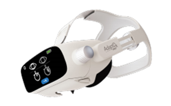
Functional testing for impaired dark adaptation not only helps to accurately det
ect AMD at a subclinical stage, it is also a tool used to monitor the disease and measure treatment effectiveness.
Several imaging tests are used to evaluate physical changes in the macula. These can detect and monitor drusen formation and changes in size, plus geographic atrophy (GA) and neovascularization in later stages.
Several tests can be administered to assess the likelihood of developing AMD. These are not diagnostic tools, but are used to identify patients with higher risk for macular degeneration.
Though useful for monitoring progression in the late stages of the disease, these tests are not considered sensitive enough to diagnose AMD at an early stage.
Diagnosing the earliest stages of AMD can present a challenge because patients are often asymptomatic, have good corrected visual acuity and the retina looks normal upon structural evaluation. Consequently, a routine vision test and structural evaluations may not raise any suspicion of the disease.
Fortunately, we now know that impaired dark adaptation function is the earliest biomarker of AMD—with impairment taking place at a subclinical level at least three years before drusen are visible. By measuring dark adaptation function in patients over age 50—especially when presenting with night vision complaints—eye care professionals can now detect subclinical AMD before any damage has been done. Once subclinical AMD is diagnosed, disease progression can be slowed through early treatment and proactive disease management.
AdaptDx Pro™ is the only test for dark adaptation that can be used quickly and effectively in a clinical setting. It uses an objective functional measure called the Rod Intercept™ (RI). Unlike traditional dark adaptation testing, the AdaptDx Pro Rapid Test takes less than 6.5 minutes. Easy to administer and interpret, it is reimbursable and detects AMD with 90.6% accuracy.
Using a single number, the Rod Intercept, you can identify dark adaptation consistent with AMD before your patient experiences irreversible vision loss.